3D Eyelid Anatomy Animation for a Dermatologic Surgeon’s Education Series
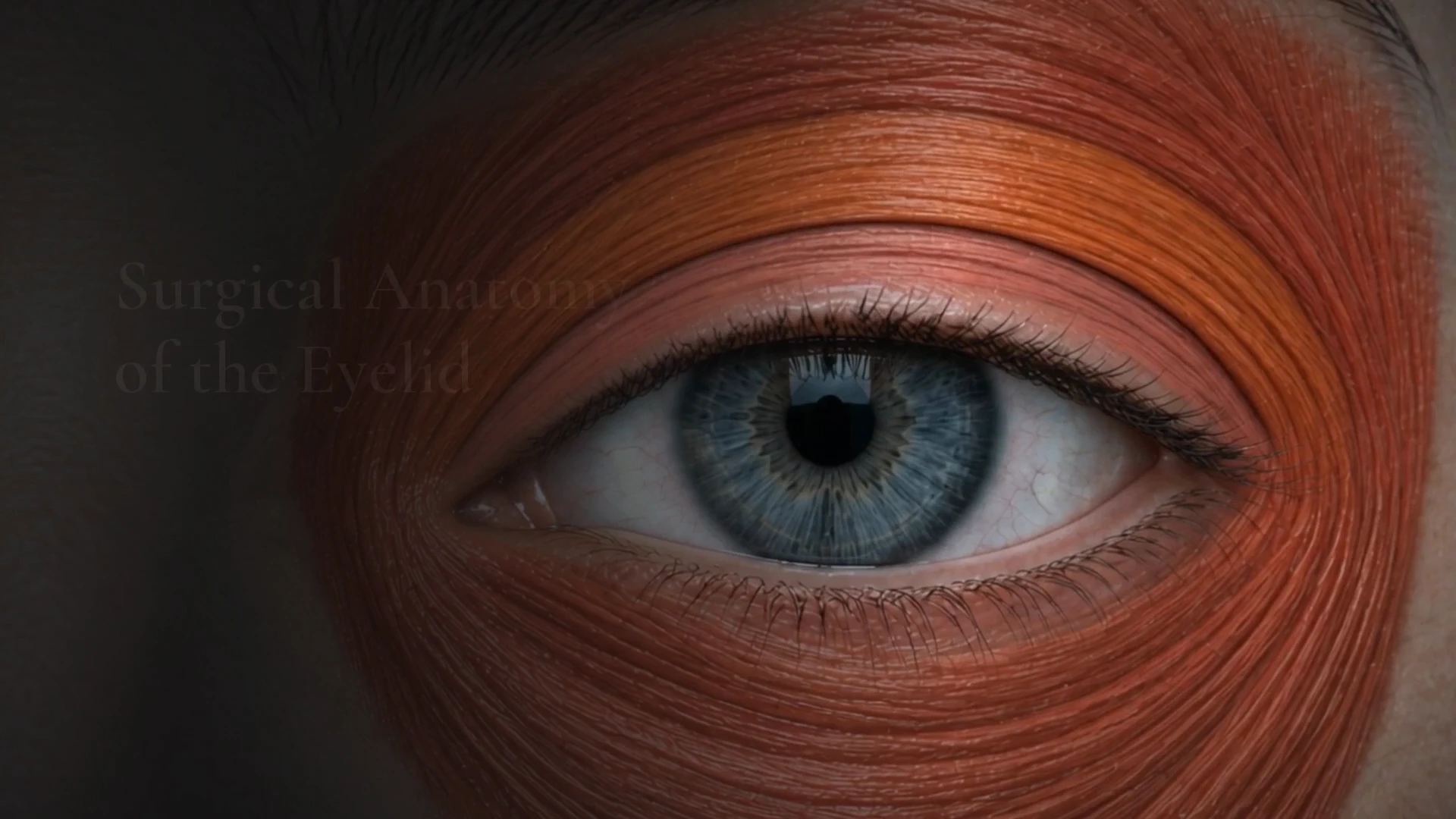
A US dermatologic surgeon needed a physician-level film of eyelid anatomy for a presentation and an education series. We storyboarded 30 shots across five parts, revised the storyboard to his notes and figures, and delivered a 5:45 3D animation timed to his narration.
Client & Market Context
A Surgeon Teaching Eyelid Anatomy to Physicians
Our client is a board-certified dermatologist and dermatologic surgeon in the United States. He needed a realistic 3D film of eyelid anatomy to narrate in a presentation for physicians and in a recorded education series.
Eyelid Layers Accurate Enough for Surgeons
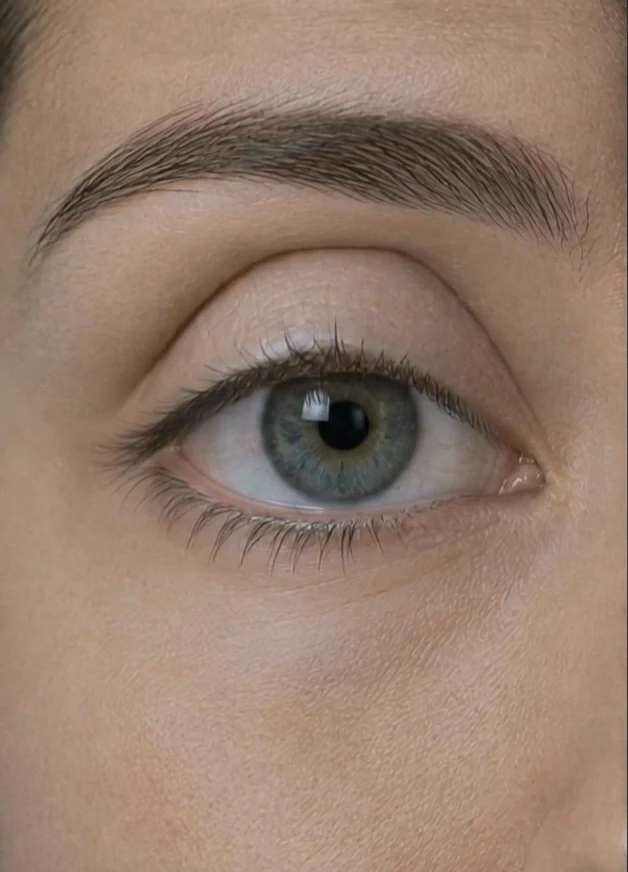
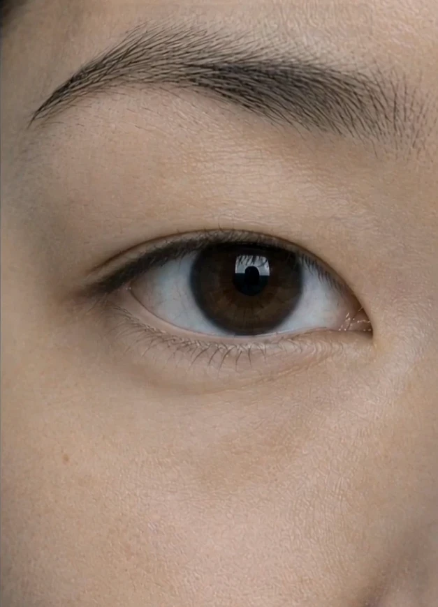
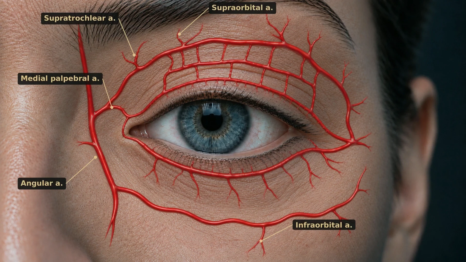
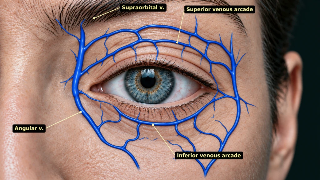
The film had to peel the lid from skin to septum to fat without losing orientation, compare two eyelid types side by side, overlay arteries, veins and nerves on the same face, and end with two procedures on the lateral canthus. Across the rounds the surgeon sent notes, figures and annotated slides, and they all had to land in one visual style.
Visual Results
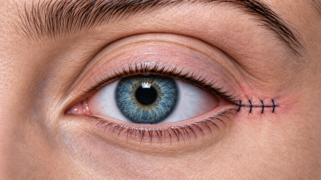
Eyelid Layers, Vessels, Nerves and Canthal Surgery
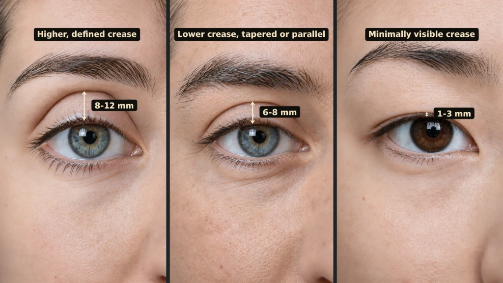
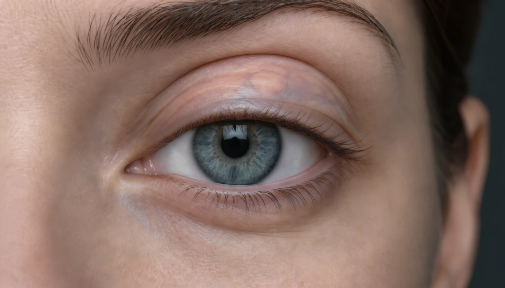
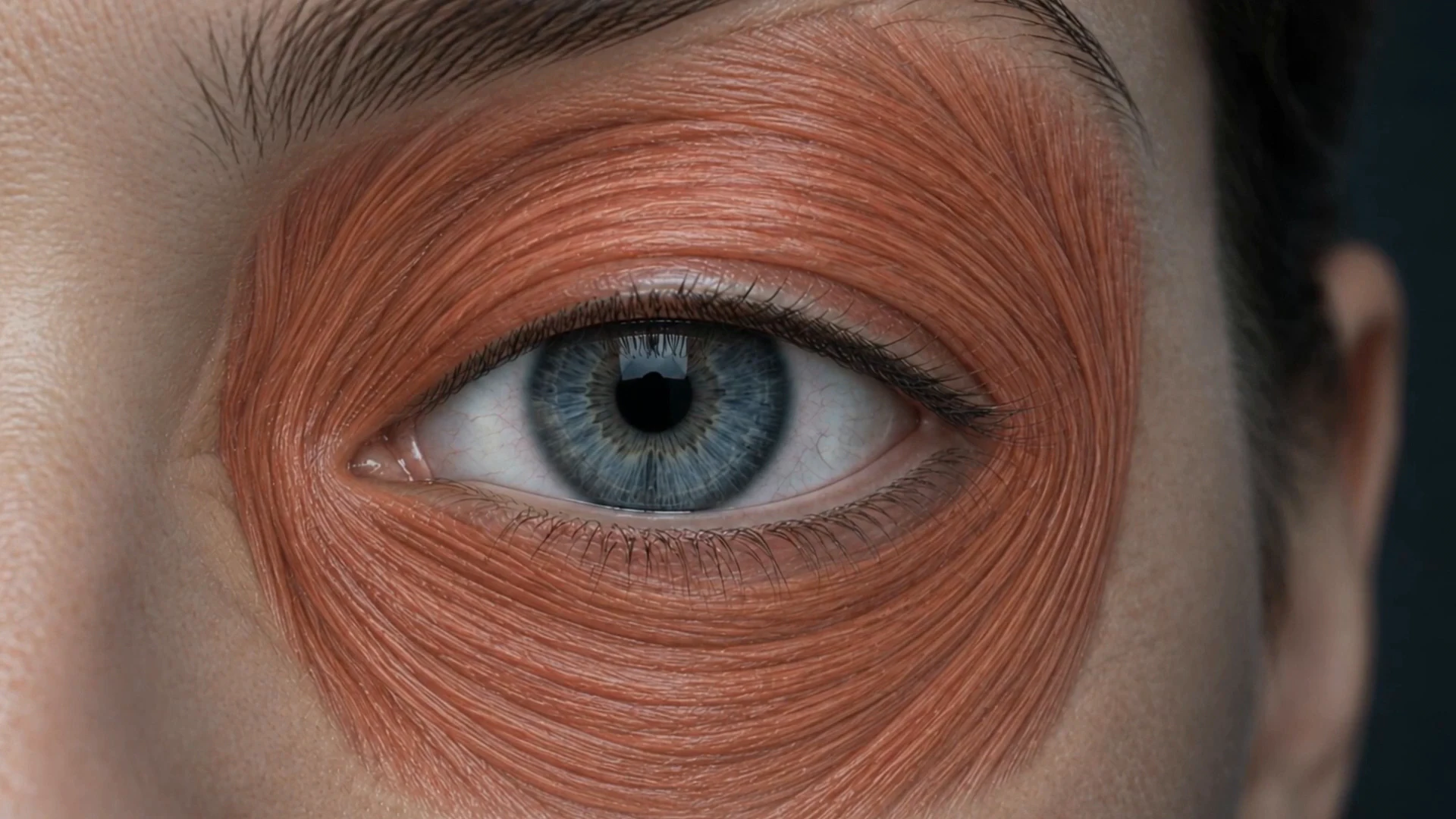
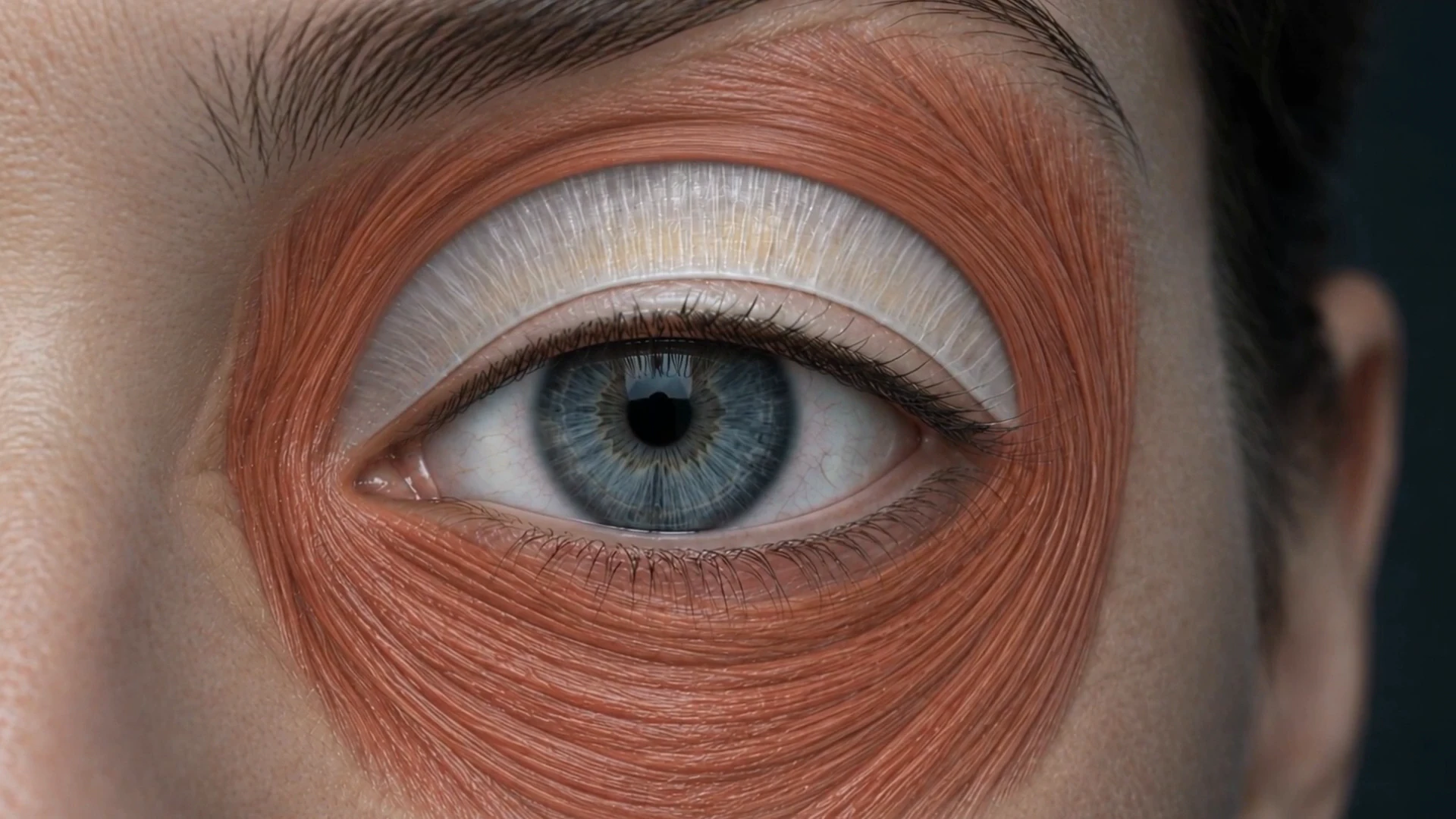
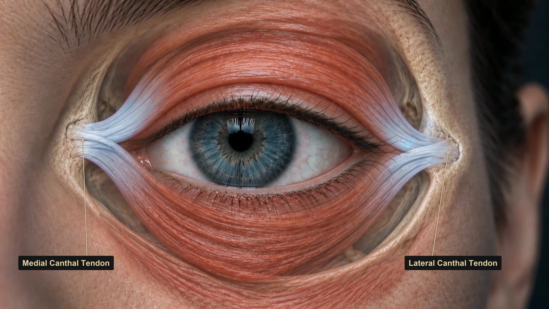
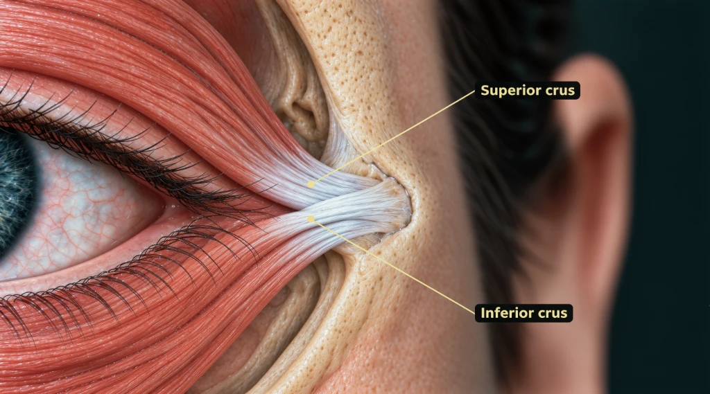
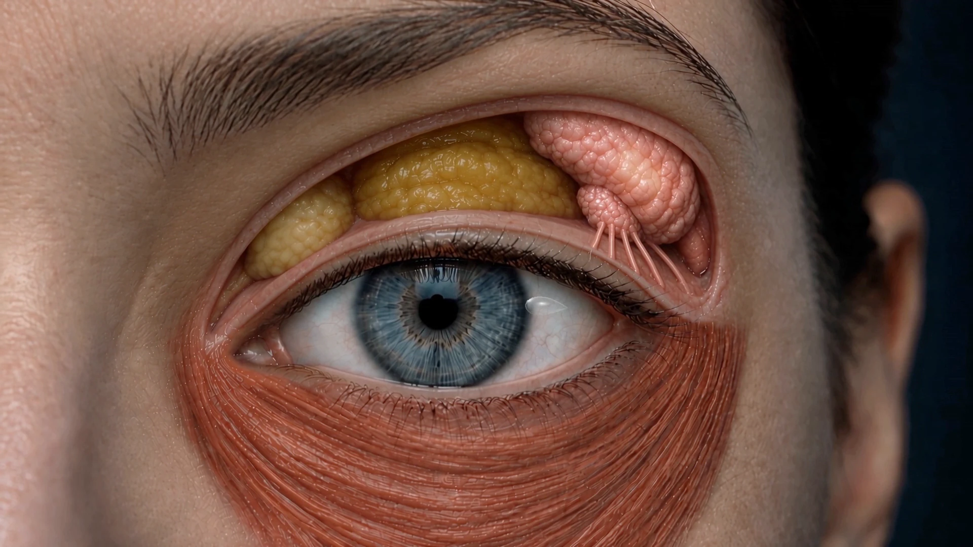
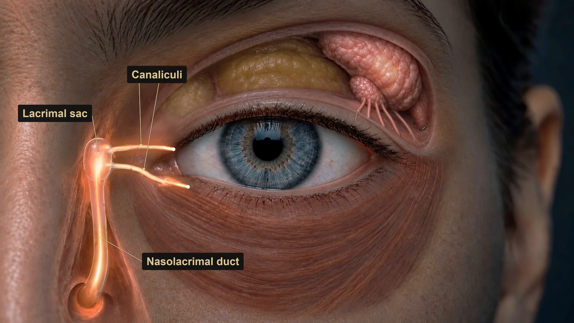
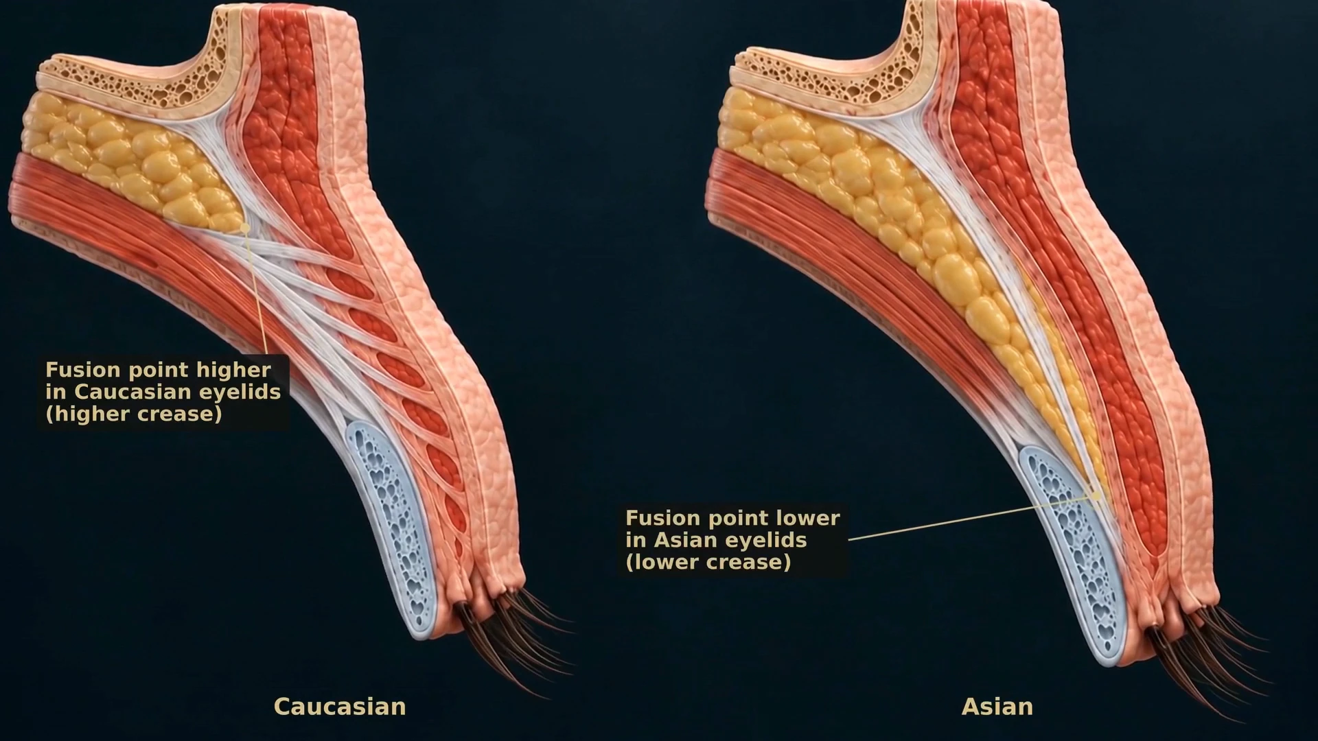
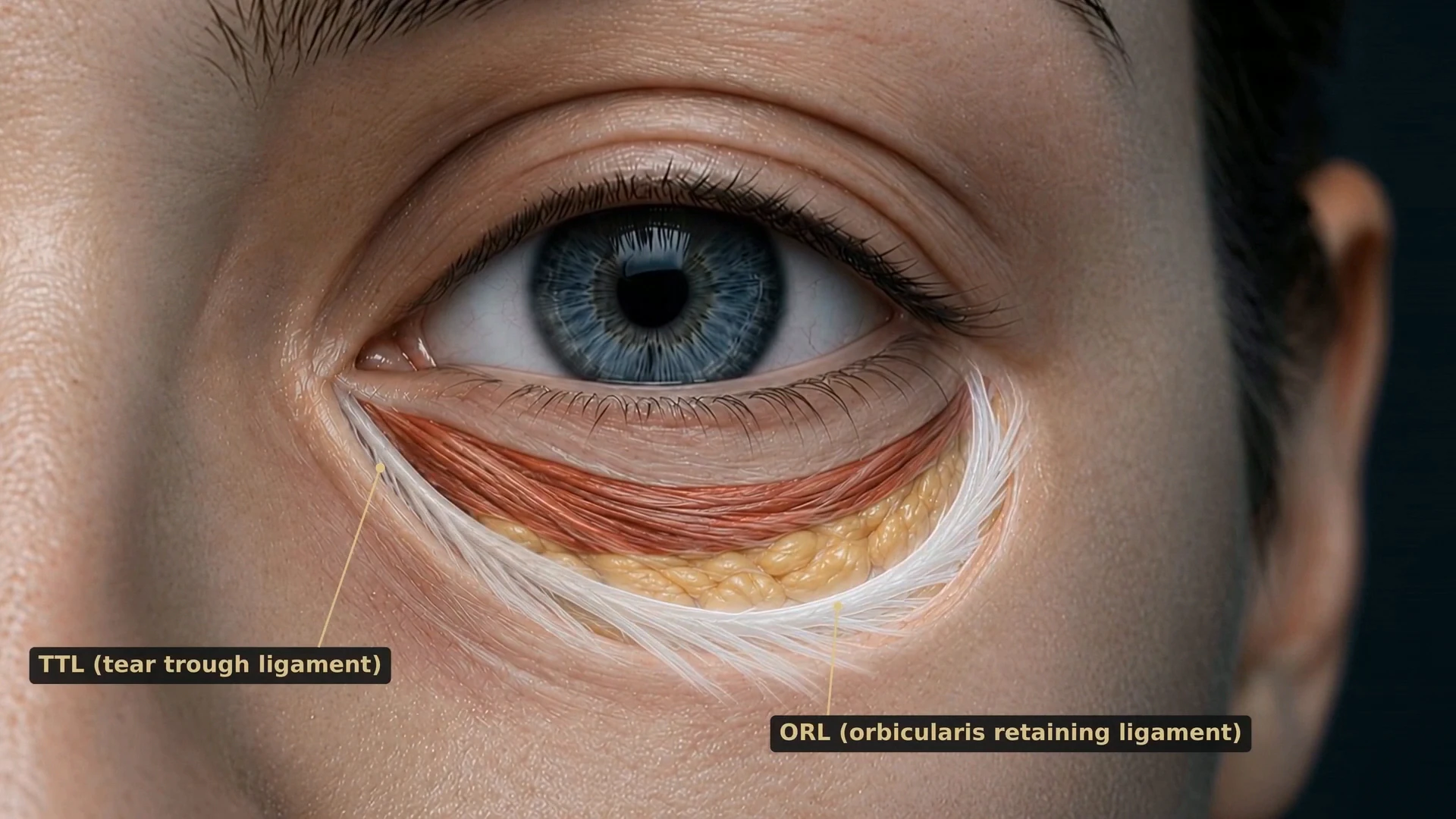
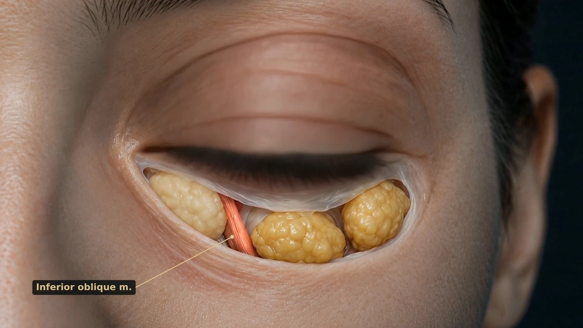
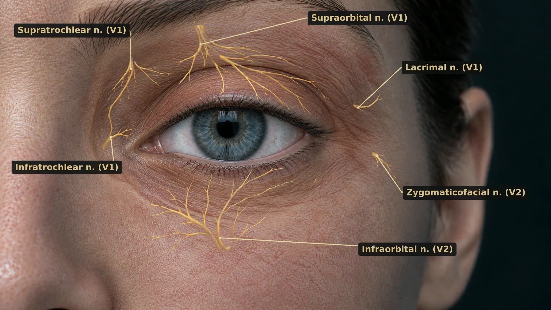
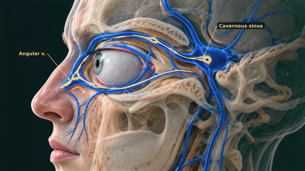
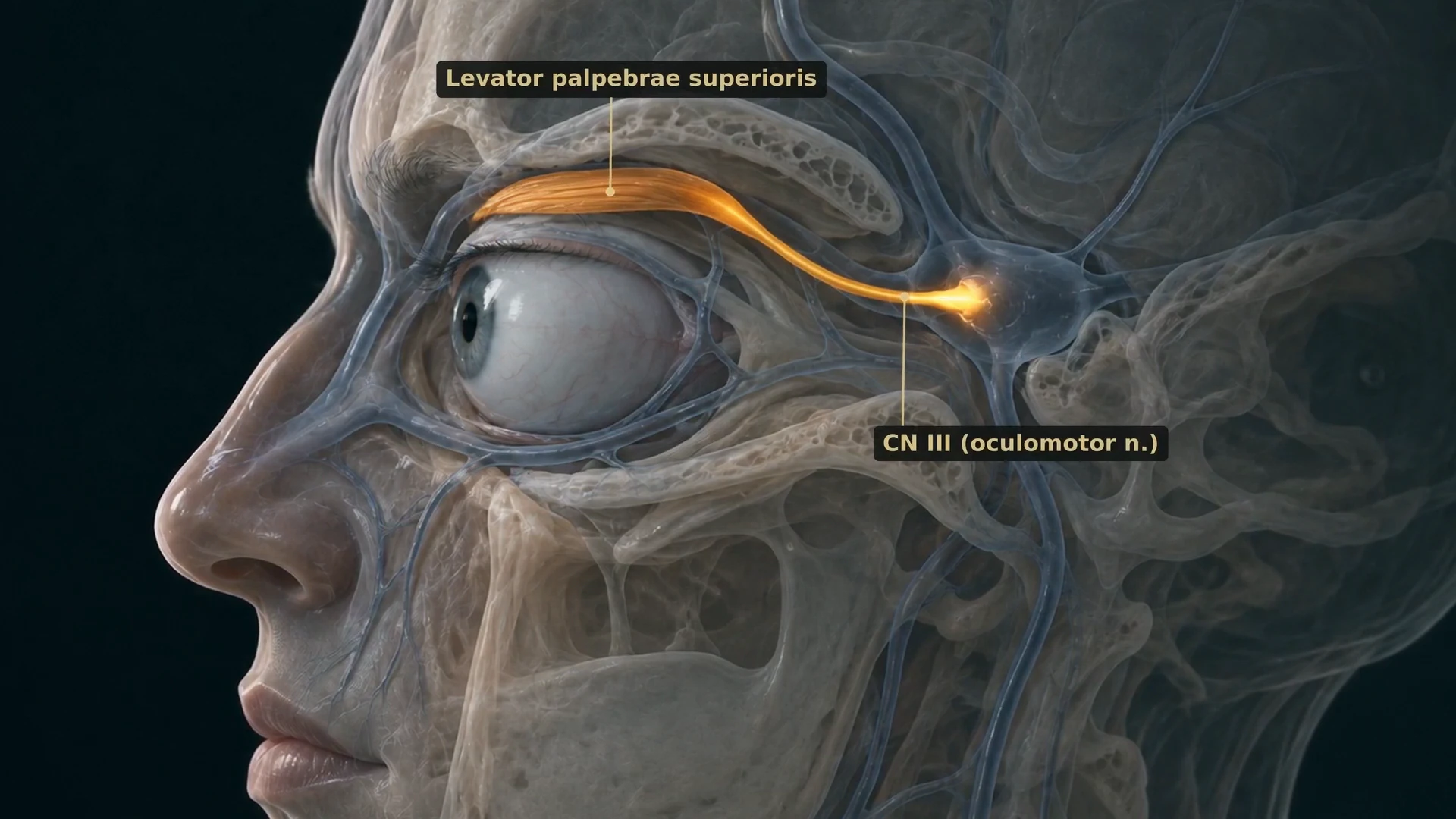
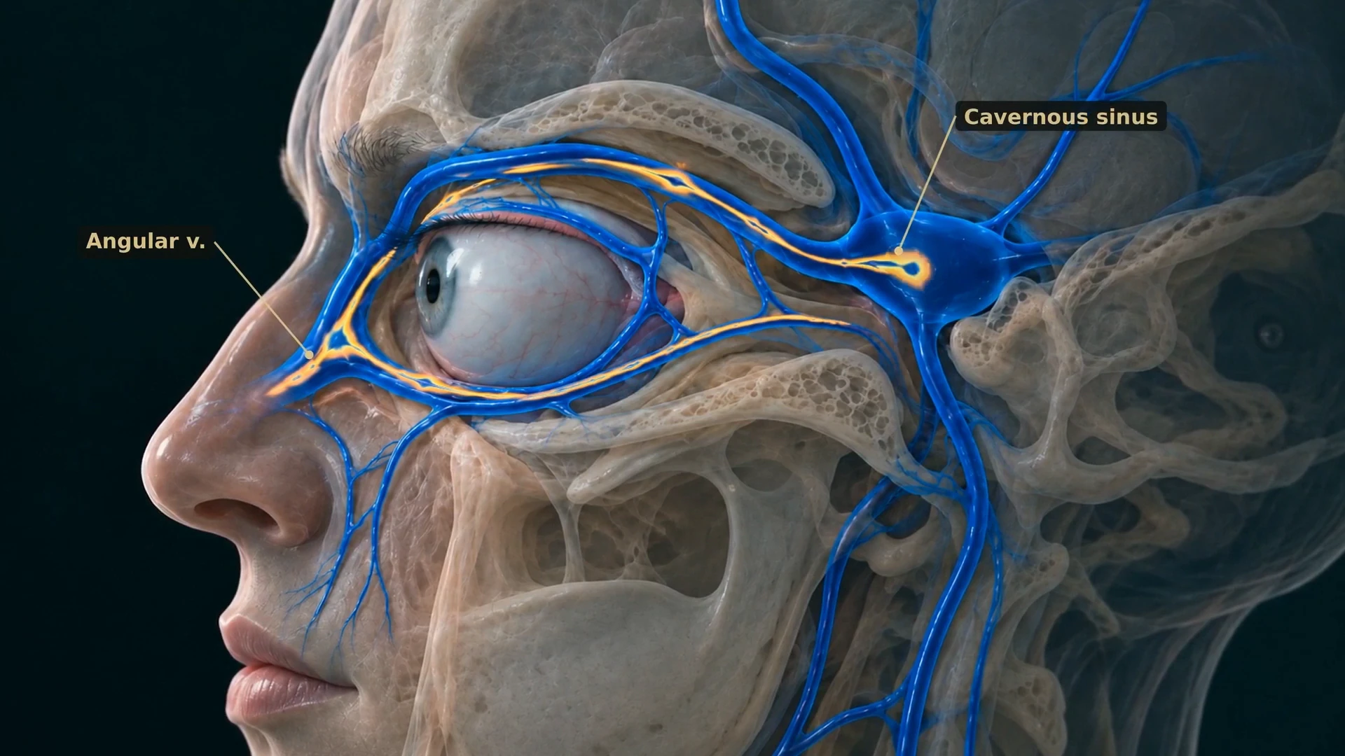
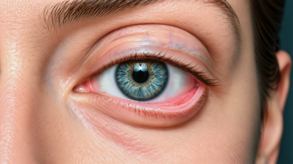
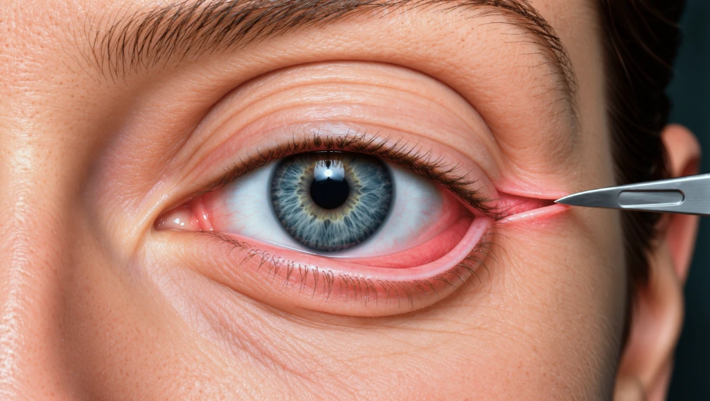
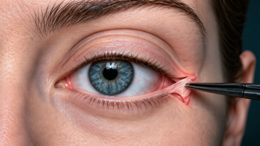
More frames from the film: the orbicularis oculi, the upper septum, tear drainage, the retaining ligaments, the venous drainage to the cavernous sinus, sensory and motor nerves, the canthal tendons and the lateral tarsal strip.
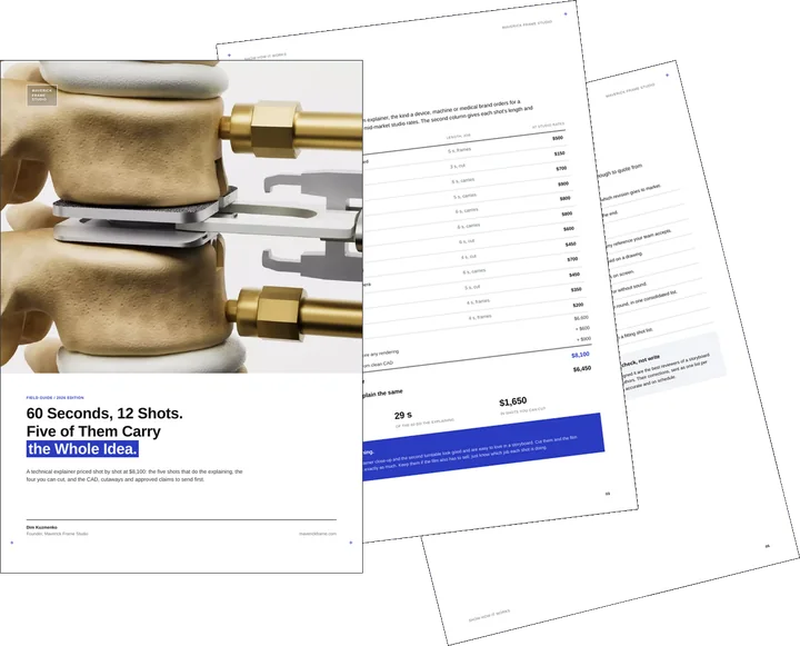
Free guide: a 60-second explainer in 12 shots, and the 4 you can cut
You have just seen a machine, a device or a procedure shown rather than described. Doing the same in motion is a matter of planning shots, not polishing renders. Our composite 60-second explainer, priced shot by shot at $8,100, does all its explaining in five shots. The guide gives you the priced shot list, the input to send from engineering or medical teams and the short brief for product animation.
- Free
- Instant download
- No sales call
If it didn't open, download it here.
Download the PDFRemote studio, your time zone
Planning a similar project?
Book a 30-minute call. Tell us about the project, and we will tell you which views it needs, how long it takes and what it costs.
Estimate on the call
A price range on the call, not a follow-up questionnaire.
Any stage of the project
From concept sketches to a finished model: we start from what you have.
Book a 30-minute call
Which day suits you?
What time?
Where should we send the invite?
- 30 minutes
- Free, no obligation
- Invite by email
Results
- 30
Shots in the approved storyboard
- 5
Parts, from the upper lid to the lateral canthus
- 5:45
Final film, timed to the surgeon’s narration
- 2
Lateral canthal procedures shown step by step
A surgical anatomy film for physicians is reviewed like a dissection: one fixed camera, one model and labels placed on the structure itself let the surgeon check every layer before the animation is rendered.
Services Provided
FAQ
FAQ About Eyelid Anatomy and Surgical Animation
Didn’t find your question?
Ask it here. A producer replies within one business day — with an answer, not a sales call.
What is an eyelid anatomy animation?
Can you animate blepharoplasty and other eyelid procedures?
How do you make sure the anatomy is accurate?
Can I narrate the animation myself?
What do you need to start a surgical anatomy animation?
How long should an anatomy animation for a lecture be?
Explore Similar Medical and Surgical Animation Cases
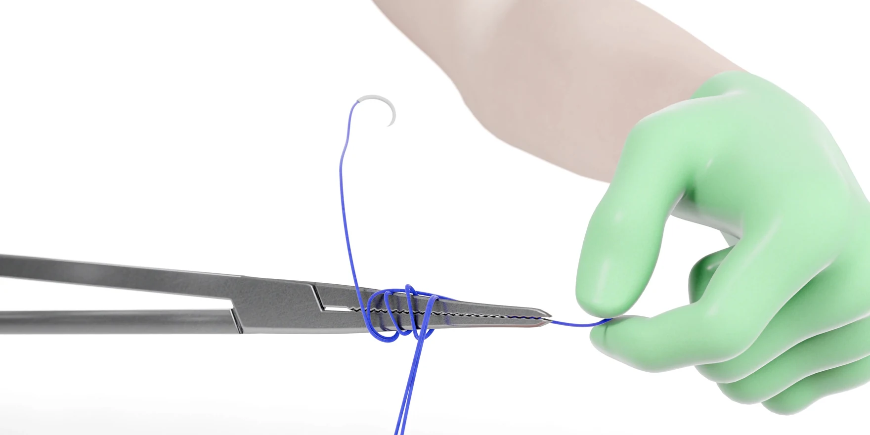
Surgical Knot Training Animation for a VR Surgery Training Company
Surgeons learn one technique at a time, and a surgical knot training animation shows the sliding knot for a VR training company with the needle driver held tip-down.
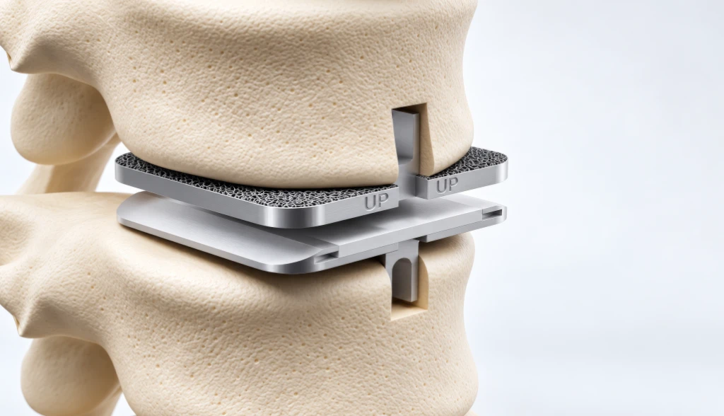
3D Surgery Animation for Medical Education with Clinical Accuracy
Procedures can be taught step by step on screen, and a 3D surgery animation for medical education turns a complex operation into doctor-approved visuals.
Medical Device Launch Video for a Clinical Eye Tracker
The eye is also a product market, and a launch film for a clinical eye tracker was built in 3D from the manufacturer’s CAD files.